Expedite production of clinically validated imaging AI algorithms with ARK
See why top healthcare systems and academic research centres around the globe trust ARK.
Schedule a DemoExperience ARK 3.0. Talk to us today!
Why Build Your Own Al Algorithms?
$ 40 Mn
Cost of diagnostic errors in Imaging annually
80,000
Deaths annually related to misdiagnosis
$ 1.8 Bn
Malpractice payouts over 10 years
AI can help identify clinical findings, save turn around time and predict likelihood of tumor recurrence and evolution from analyzing diagnostic imaging.
Create Your Own Al To Detect Critical Anomalies For All Body Parts And Abnormalities
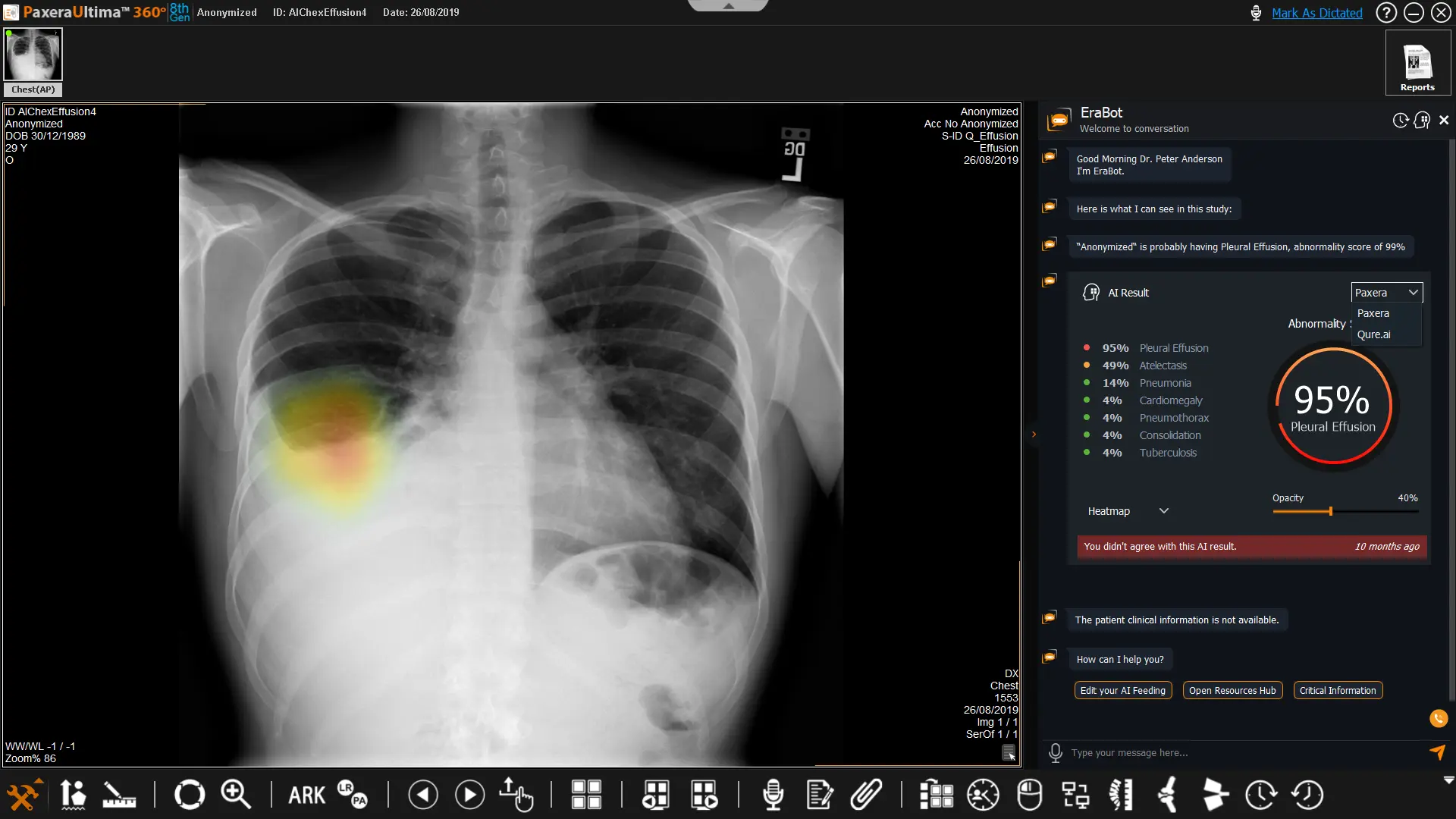
Robust Viewer
A robust zero-footprint viewer offers on-demand accessibility to image analysis tools and multi-specialty measurements. Its user-friendly interface facilitates the creation of powerful data annotations and the establishment of model ground truth.
Furthermore, it provides the benefit of strong interoperability by seamlessly integrating with various PACS, VNA, and research archives. This integration enhances workflow efficiency through the exchange of presentation states and the chatbot.
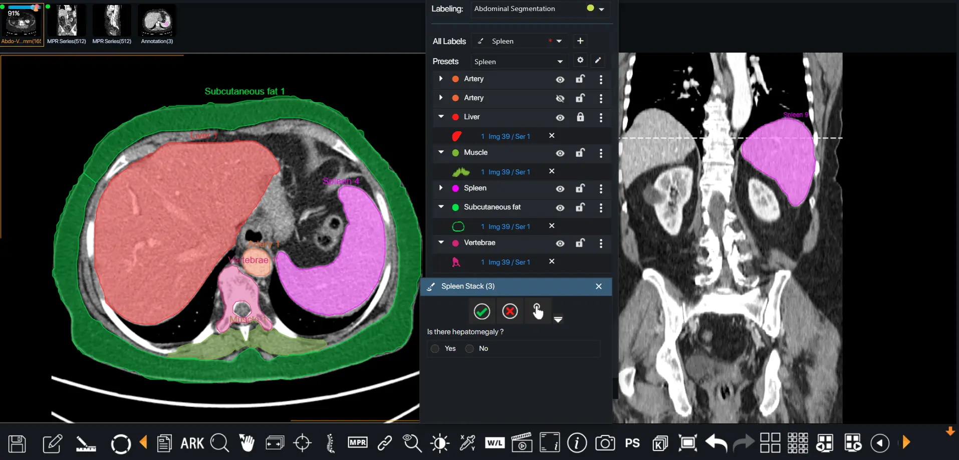
Data Curation and Segmentation
Leverage your data annotations to derive meaningful radiomics insights and enhance your segmentations with labels and descriptions. Conduct data quality checks to identify and remove erroneous samples, ensuring optimal model training.
Ark offers advanced techniques for both manual and automatic segmentation, which can be exported in various formats.
AI Modeling
Make ML modeling a breeze! Select the best architecture for your data from our diverse model library or build a custom AI model from scratch. Fine-tune and control different versions of your model.
Boost your model’s training and predictability by integrating your clinical data points and harnessing data from multi-modal sources.
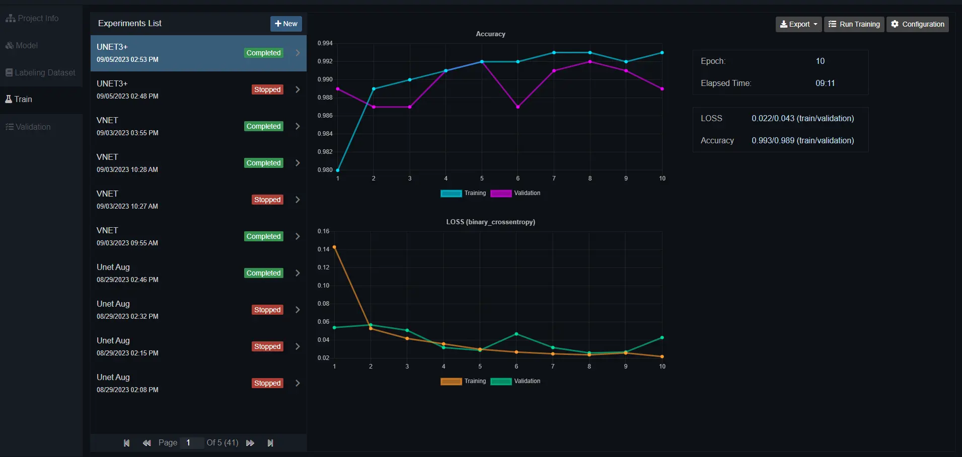
Training and Validation
Create as many training experiments as necessary and gain complete visibility into the model’s performance as training progresses.
Take advantage of insightful charts to fine-tune your hyperparameters.
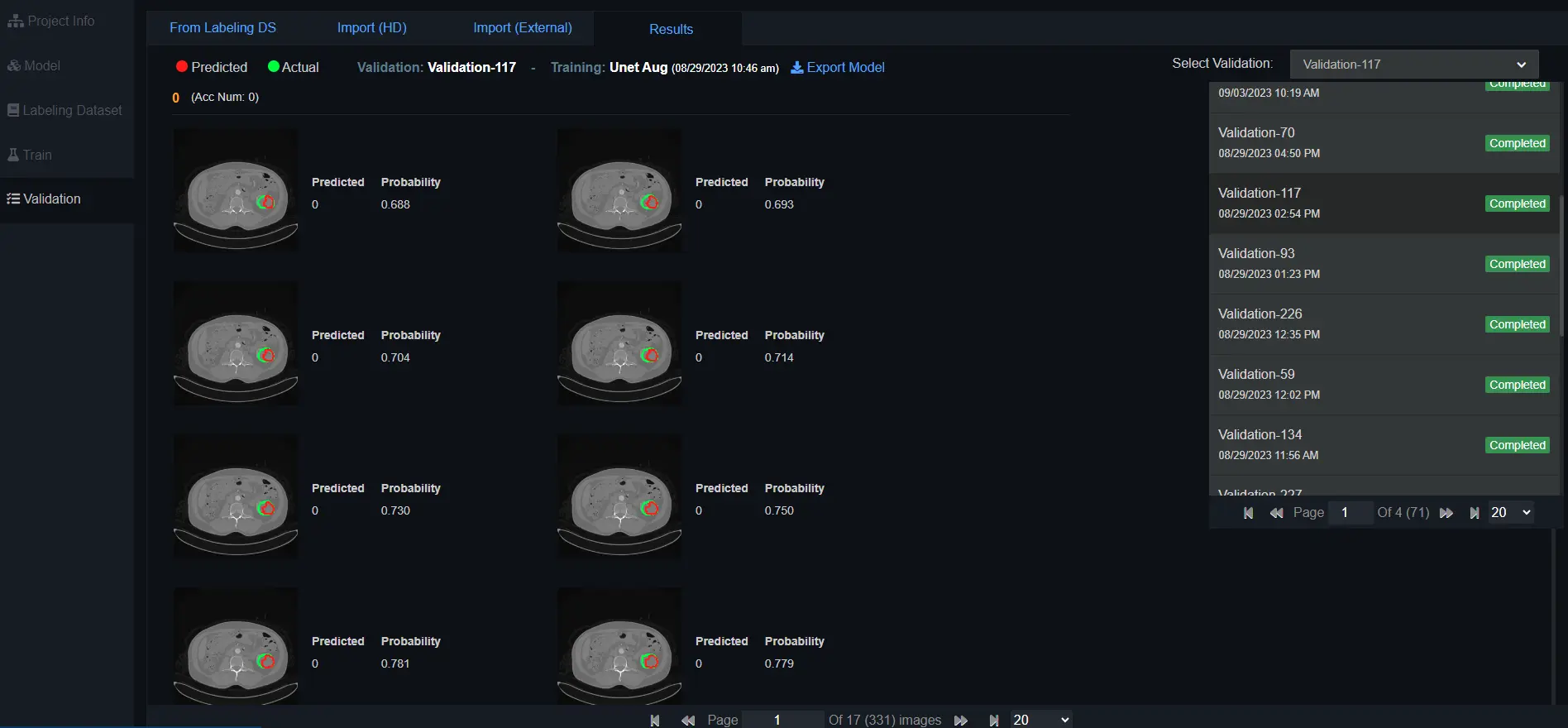
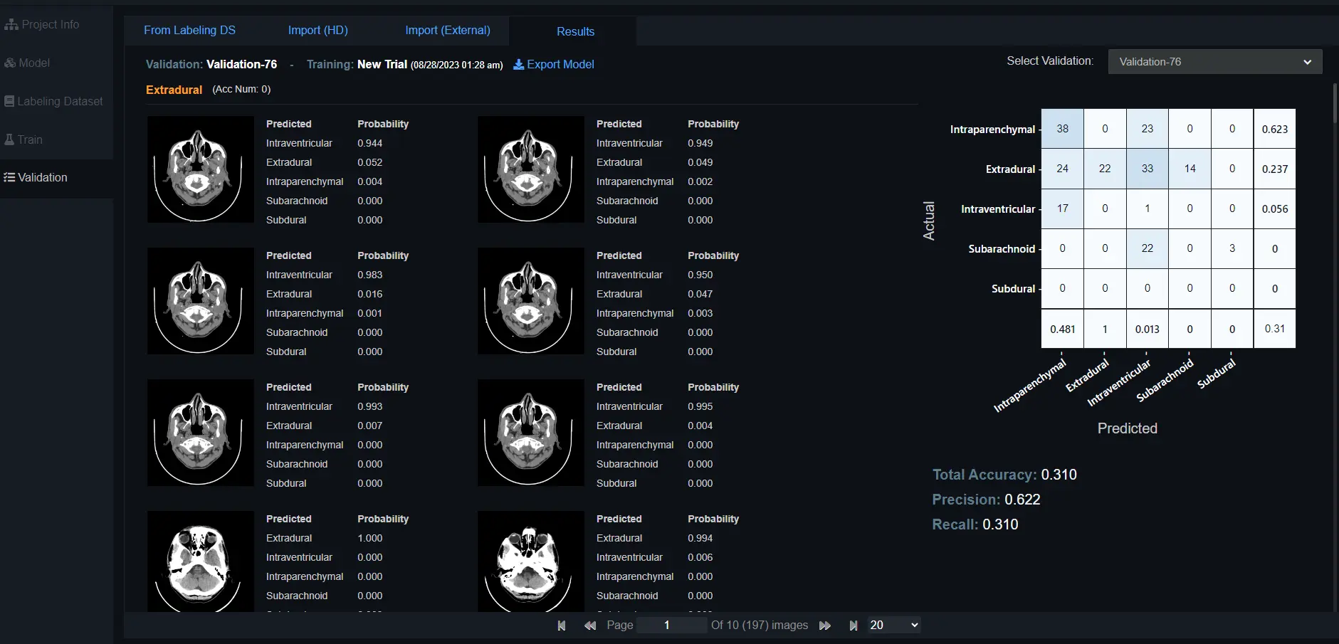
Testing and Results
Test and cross-validate your model’s performance on diverse subsets of data to ensure its ability to generalize to new data points.
Gain deeper insights into the model’s prediction results and confidence levels well in advance.
Identify misclassifications early with powerful binary and multi-class confusion matrix results.
Additionally, export your algorithms into Docker containers for seamless integration into your clinical workflow.
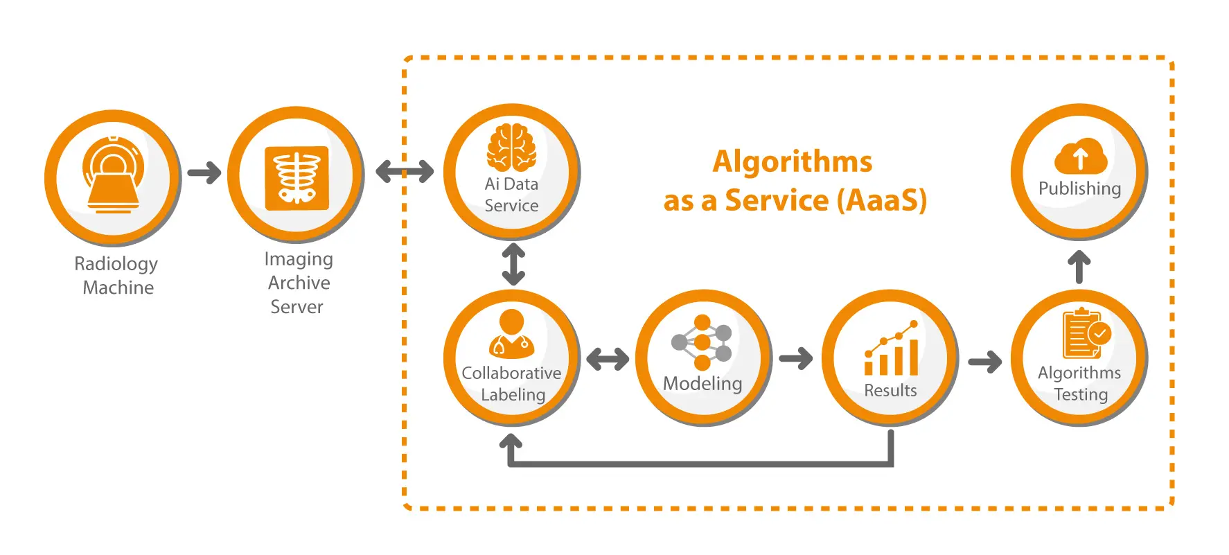
Algorithms as a Service
Our mission is to help reduce diagnostic errors in medical imaging by democratizing our AI, making it accessible, affordable, safer and faster to deploy, thus reducing overall healthcare costs. We provide a full suite of authoring tools with an unbiased dataset to help users self-develop AI/ML algorithms from start to finish.
Zero Coding
We provide a Do-It-Yourself (DIY) authoring platform that requires no coding to produce AI algorithms for all possible modalities, body parts, and anomalies. With high-end modeling, annotation, and native data curation tools, users are empowered to securely self-develop algorithms quickly and without extensive coding.
Accessible, Affordable, and Safer Algorithm Creation and Deployment
Board the Ark Today